Simulation technologies offer interesting opportunities for computer planning of orthognathic surgery. However, the methods used to date require tedious set up of simulation meshes based on patient imaging data, and they rely on complex simulation models that require long computations. In this work, we propose a modeling and simulation methodology that addresses model set up and runtime simulation in a holistic manner. We pay special attention to modeling the coupling of rigid-bone and soft-tissue components of the facial model, such that the resulting model is computationally simple yet accurate. The proposed simulation methodology has been evaluated on a cohort of 10 patients of orthognathic surgery, comparing quantitatively simulation results to post-operative scans. The results suggest that the proposed simulation methods admit the use of coarse simulation meshes, with planning computation times of less than 10 seconds in most cases, and with clinically viable accuracy.
Keywords: Soft-tissue simulation; finite-element model; surgical planning; orthognathic surgery@Article{jpm11100982,
AUTHOR = {Alcañiz, Patricia and Pérez, Jesús and Gutiérrez, Alessandro and Barreiro, Héctor and Villalobos, Ángel and Miraut, David and Illana, Carlos and Guiñales, Jorge and Otaduy, Miguel A.},
TITLE = {Soft-Tissue Simulation for Computational Planning of Orthognathic Surgery},
JOURNAL = {Journal of Personalized Medicine},
VOLUME = {11},
YEAR = {2021},
NUMBER = {10},
ARTICLE-NUMBER = {982},
URL = {https://www.mdpi.com/2075-4426/11/10/982},
ISSN = {2075-4426},
ABSTRACT = {Simulation technologies offer interesting opportunities for computer planning of orthognathic surgery. However, the methods used to date require tedious set up of simulation meshes based on patient imaging data, and they rely on complex simulation models that require long computations. In this work, we propose a modeling and simulation methodology that addresses model set up and runtime simulation in a holistic manner. We pay special attention to modeling the coupling of rigid-bone and soft-tissue components of the facial model, such that the resulting model is computationally simple yet accurate. The proposed simulation methodology has been evaluated on a cohort of 10 patients of orthognathic surgery, comparing quantitatively simulation results to post-operative scans. The results suggest that the proposed simulation methods admit the use of coarse simulation meshes, with planning computation times of less than 10 seconds in most cases, and with clinically viable accuracy.},
DOI = {10.3390/jpm11100982}
}
The proposed simulation methodology has been evaluated on a cohort of 10 patients of orthognathic surgery, comparing quantitatively simulation results to post-operativescans. We collected the following data for each patient: age (mean 32 years, range 22–51 years), gender (8 women, 2 men), ethnic group (8 Caucasian, 2 Latin American), and diagnosis (2 class II malocclusion cases, 4 class III malocclusion cases, 3 asymmetry cases, 1 open bite case). The surgeries undergone by the patients exhibit diverse procedures for both the maxilla and the mandible:
• In maxillary procedures, the maxilla is separated from the skull through a Lefort osteotomy, classified based on its anatomical level. In this cohort, the distribution of cases is: 8 Lefort I cases and 1 Lefort II case; one patient did not undergo maxillary surgery. Moreover, after a Lefort I osteotomy, the maxilla may be segmented (typically into three fragments) in order to expand the upper arch. Maxilla segmentation was applied to 6 patients in this cohort.• In mandibular procedures, the mandible may be sagittally split on both rami (bilateral sagittal split osteotomy, BSSO) or only one ramus (unilateral sagittal split osteotomy, USSO). In this cohort, the distribution of cases is: 7 BSSO cases, 1 USSO case; two patients did not undergo mandibular surgery. Additionally, a chin osteotomy or genioplasty may be also performed. Genioplasty was applied to 1 patient in this cohort.


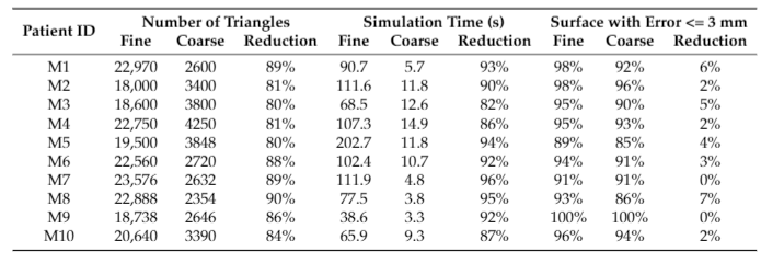
The cumulative error analysis summarized in Table 2 indicates a small loss of accuracy when the resolution of the simulation meshes is reduced. On average, 92% of the surface of the patients has an error lower than 3 mm with coarse meshes, and with fine meshes this percentage grows to 95%. Simulations with coarse meshes also exhibit a slightly wider range of error values. However, when the specific patient cases are inspected in more detail, as depicted in Figures 1 and 2, we can see that error appears in the same areas with coarse and fine meshes. The use of coarse meshes does not lead to additional sources of error, and the coarse and fine simulations are qualitatively equivalent. The accuracy of coarse simulation meshes indicates that for most clinical cases they are perfectly valid, as the error in critical areas remains under clinically acceptable thresholds (i.e., 3 mm). In the worst case, the coarse simulation can be used as a faster preview of the clinical prediction, which can dramatically accelerate planning iterations. Only when the coarse simulation provides a clinically satisfactory result, surgeons may launch a fine simulation for higher accuracy. The combination of coarse and fine simulations is further justified by the extreme reduction in computation times. As listed in Table 2, the reduction in simulation times achieved with coarse meshes (90.7% on average) is higher than the reduction in mesh complexity (84.8% on average). Moreover, this drastic reduction in simulation times produces only a minimal reduction in simulation accuracy (3.1% on average, measured as the cumulative surface percentage with error below 3 mm).

The analysis of results suggests that coarse meshes are accurate enough for full prediction of the clinical intervention in some cases. In other cases, due to the slight increase in error, we advise executing a final prediction using fine meshes. The use of coarse meshes can anyway have a strong impact in practical planning situations, as clinicians will be able to execute fast planning using coarse meshes as a good preview of the final result.